Hypervascular subtype is characterised by the presence of regressed germinal centres and follicular dendritic cell prominence, whereas the plasmacytic subtype exhibits hyperplastic germinal centres and profuse plasmacytosis.
HYPERVASCULAR SUBTYPE
MIXED SUBTYPE
PLASMACYTIC SUBTYPE
EXPERT GUIDANCE
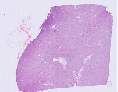
Plasmacytic subtype
This lymph node is characterised by some degree of follicular hyperplasia with hyperplastic GCs (Grade 1), but no regression of GCs (Grade 0) or FDC prominence (Grade 0). There is prominent interfollicular plasmacytosis (Grade 3). There is no increased vascularity (Grade 0).
Abbreviations
GCs, germinal centres; FDC, follicular dendritic cell.
This tool has been funded and produced by Recordati. All images have been provided by an international panel of expert pathologists. The concept, functionality and expert guidance found within this tool has also been developed with the support of expert pathologists.
EXPERT GUIDANCE
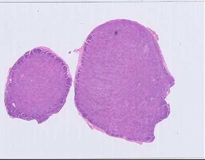
Mixed subtype
This lymph node is characterised by a mixture of features. There is regression of GCs (Grade 2), FDC prominence (Grade 2), and increased vascularity (Grade 2), combined with an interfollicular plasmacytosis (Grade 2). In the periphery of the lymph node there is some follicular hyperplasia with hyperplastic GCs (Grade 1).
Abbreviations
GCs, germinal centres; FDC, follicular dendritic cell.
This tool has been funded and produced by Recordati. All images have been provided by an international panel of expert pathologists. The concept, functionality and expert guidance found within this tool has also been developed with the support of expert pathologists.
EXPERT GUIDANCE
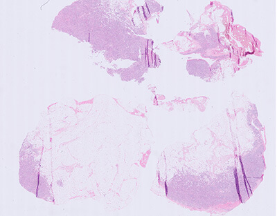
Hypervascular subtype
This lymph node is characterised by prominent regression of the majority of GCs (Grade 3). There is prominent vascularity with hyalinisation in the majority of the GCs (Grade 3). There is no follicular hyperplasia with hyperplastic GCs (Grade 0). There is an increase of high endothelial venules in the interfollicular space. There is a moderate number of plasma cells (Grade 1).
Abbreviations
GCs, germinal centres; FDC, follicular dendritic cell.
This tool has been funded and produced by Recordati. All images have been provided by an international panel of expert pathologists. The concept, functionality and expert guidance found within this tool has also been developed with the support of expert pathologists.
You have clicked an external link and are now leaving
this non-product Recordati website